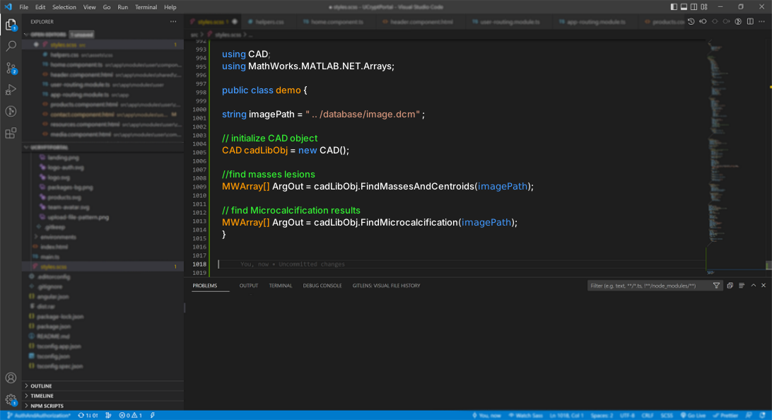
LIBCAD is a detection engine that can be integrated into other software or accessed through an API. WEBCAD is the browser-based interface built around it.
The system combines mammogram processing with several ways to inspect or integrate its output.
01
Machine-independent processing
The filtering method is designed to reduce dependence on machine brand, resolution, and acquisition characteristics.
02
Several deployment forms
LIBCAD can be provided as a Linux shared object, a Windows dynamic-link library, or a remotely accessed API.
03
Several output types
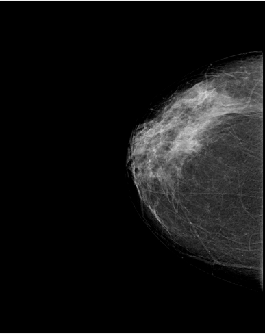
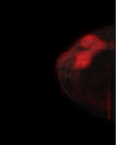
Outputs include mass scores, markers, microcalcification results, filtered images, and enhanced images.
04
CPU and GPU processing
Parts of the system use parallel processing and hardware acceleration.
05
Software integration
The engine is intended for integration into research and imaging-software workflows.
06
Research foundation
The system grew from published work in mammography analysis and computer-aided detection.
Research record
Patent and selected publications.
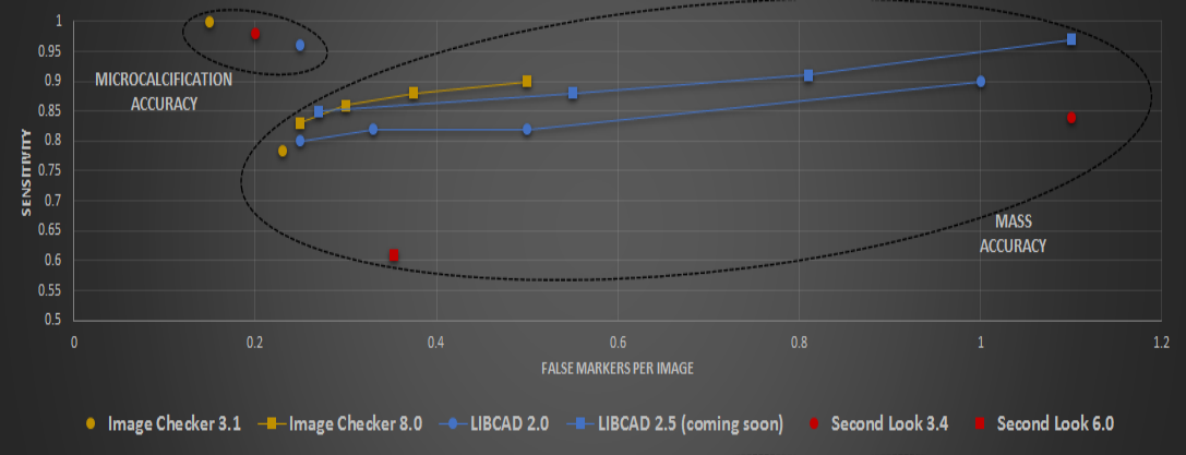
The results below belong to the research record. They are not presented as a current clinical-performance claim.
U.S. patent 1
Yousef, W. A. “Method and system for computer aided detection for cancer.” U.S. Patent 10,789,712, September 2020.
Selected publications 4
Yousef, W. A. et al. (2019). “Method and System for Image Analysis to Detect Cancer.” arXiv:1908.10661.
Abdel Razek, N. M., Yousef, W. A., & Mustafa, W. A. (2013). “Microcalcification detection with and without CAD system (LIBCAD): A comparative study.” Egyptian Journal of Radiology and Nuclear Medicine, 44(2), 397–404.
Abdelrazek, N., Yousef, W., & Mustafa, W. (2012). “Microcalcification detection with and without prototype CAD system (LIBCAD): a comparative study.” ECR 2012 / C-1063.
Yousef, W. A. et al. (2010). “On Detecting Abnormalities in Digital Mammography.” 39th Applied Imagery Pattern Recognition Workshop.
Reported performance chart Research resultComparison retained from the existing LIBCAD research materials.LIBCAD manual By request
Email MESC Labs for the current manual and to discuss the appropriate integration form.
Static demonstration
One demonstration page, two clearly separated case series.
No images are uploaded, no new analysis is performed, and nothing shown here should be used for clinical decisions.
01
Comprehensive output explorer
Compare original and processed views across eight cases.
Inspect mass scores, markers, microcalcifications, and enhancement levels beside the original mammograms. A separate ten-case annotated enhancement series appears farther down the same page.